Atjaunināt sīkdatņu piekrišanu

Dermatoscopy and Skin Cancer, second edition: A handbook for hunters of skin cancer and melanoma [Mīkstie vāki]

  • Formāts: Paperback / softback, 384 pages, height x width x depth: 244x172x17 mm, weight: 975 g
  • Izdošanas datums: 31-Aug-2025
  • Izdevniecība: Scion Publishing Ltd
  • ISBN-10: 1914961676
  • ISBN-13: 9781914961670
Citas grāmatas par šo tēmu:
  • Mīkstie vāki
  • Cena: 61,21 €
  • Grāmatu piegādes laiks ir 3-4 nedēļas, ja grāmata ir uz vietas izdevniecības noliktavā. Ja izdevējam nepieciešams publicēt jaunu tirāžu, grāmatas piegāde var aizkavēties.
  • Daudzums:
  • Ielikt grozā
  • Piegādes laiks - 4-6 nedēļas
  • Pievienot vēlmju sarakstam
  • Formāts: Paperback / softback, 384 pages, height x width x depth: 244x172x17 mm, weight: 975 g
  • Izdošanas datums: 31-Aug-2025
  • Izdevniecība: Scion Publishing Ltd
  • ISBN-10: 1914961676
  • ISBN-13: 9781914961670
Citas grāmatas par šo tēmu:
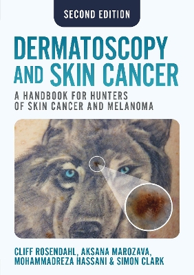
Dermatoscopy and Skin Cancer, second edition, is a handbook to help dermatologists, dermatoscopists and GPs easily differentiate between benign and malignant tumours, leading to fewer unnecessary biopsies and earlier treatment of cancers.


Based around two easy to follow algorithms, 'Chaos and Clues' and 'Prediction without Pigment', the book shows all dermatoscope users how to confidently diagnose skin lesions earlier and with greater precision.


In addition, this handbook provides coverage of:


the microanatomy of the skin specimen processing and histopathology the language of dermatoscopy to help name and define structures and patterns approaches to skin examination and photodocumentation revised pattern analysis as an additional diagnostic algorithm dermatoscopic features of common and significant lesions.


Using hundreds of high quality images, the authors provide a detailed algorithmic approach to assessing the skin; an approach that has been successfully taught to thousands of doctors around the world.


From Doody's reviews, December 2023

"Many dermoscopy books exist; some are too pedantic and explain concepts with dermatoscopic jargon, while others purport to simplify the learning process but quickly succumb to the same criticism. Most are replete with abnormal looking lesions, but fall short on including examples of normal variations. This book delivers what it promises. I definitely recommend it as the first reference for mastering diagnosis of skin lesions with a dermatoscope." 4 stars!
Preface to the second edition; Foreword; Abbreviations


Chapter 1: Introduction to dermatoscopy


1.1 Why use a dermatoscope?

1.2 What is a dermatoscope?

1.3 Colours in dermatoscopy

1.4 Differences between polarised and non-polarised dermatoscopy

1.5 Uses of dermatoscopy for conditions other than tumours


Chapter 2: Skin the organ


2.1 Skin as an organ

2.2 Embryology of skin

2.3 The microanatomy of skin


Chapter 3: Dermatopathology for dermatoscopists


3.1 Introduction

3.2 Tissue and context: the two pillars of reliable skin cancer diagnosis

3.3 From the scalpel to the microscope: the specimen journey

3.4 The histology of normal skin

3.5 Core dermatopathology terminology

3.6 Neoplastic lesions: dermatoscopichistological correlation

3.7 Clinicopathological collaboration in skin cancer diagnosis


Chapter 4: The language of dermatoscopy: naming and defining structures and
patterns


4.1 The evolution of terminology for dermatoscopic structures and patterns

4.2 Revised pattern analysis of lesions pigmented by melanin

4.3 Patterns in revised pattern analysis

4.4 The process of revised pattern analysis

4.5 Revised pattern analysis applied to lesions with white structures

4.6 Revised pattern analysis applied to lesions with orange, yellow and
skin-coloured structures

4.7 Revised pattern analysis a diagnostic algorithm

4.8 An aide-memoire for revised pattern analysis of pigmented skin lesions

4.9 Revised pattern analysis applied to vessel structure and patterns

4.10 The cognition of dermatoscopy


Chapter 5: The skin examination


5.1 The skin check consultation

5.2 Photo-documentation

5.3 Patient safety: tracking specimens and self-audit

5.4 The lives of lesions


Chapter 6: Chaos, Clues and Exceptions: a decision algorithm for pigmented
skin lesions


6.1 'Chaos, Clues and Exceptions'

6.2 Chaos

6.3 Clues

6.4 Exceptions


Chapter 7: Prediction without Pigment: an algorithm for non-pigmented skin
lesions


7.1 'Prediction without Pigment'

7.2 'Prediction without Pigment': Part 1

7.3 'Prediction without Pigment': Part 2

7.4 Conclusion


Chapter 8: Melanoma


8.1 What is a melanoma?

8.2 Melanoma subtypes

8.3 Melanomas with adverse outcomes

8.4 Metastatic melanoma


Chapter 9: Melanocytic naevi


9.1 Melanocytic naevi, pigmented and non-pigmented

9.2 Dysplastic naevus


Chapter 10: Basal cell carcinoma, benign and malignant keratinocytic lesions,
distinguishing flat pigmented facial lesions, dermatofibroma, vascular and
other lesions


10.1 Basal cell carcinoma: pigmented and non-pigmented

10.2 Benign keratinocytic lesions

10.3 Actinic keratosis and squamous cell carcinoma in situ

10.4 Squamous cell carcinoma and keratoacanthoma

10.5 Distinguishing flat pigmented facial lesions

10.6 Dermatofibroma and dermatofibrosarcoma protuberans

10.7 Haemangioma and other vascular lesions

10.8 Merkel cell carcinoma

10.9 Atypical fibroxanthoma

10.10 Adnexal tumours

10.11 Neurofibroma

10.12 Molluscum contagiosum

10.13 Cutaneous lymphoma

10.14 Kaposi sarcoma


Chapter 11: Photographic technology as a diagnostic tool in melanoma
management


11.1 Utilisation of photographic technology in skin lesion diagnostics

11.2 Serial digital dermatoscopic imaging

11.2.1 Serial digital dermatoscopic imaging targeted and random

11.2.2 Selection by serial digital dermatoscopic imaging a promising
alternative to random lesion selection (SDDI TBPD)

11.2.3 Serial digital dermatoscopic imaging which lesions should be
excised?

11.3 Total body photography

11.3.1 Total body photography used as a baseline during skin examination

11.3.2 Serial total body photography with detection software


Index