This update of the popular
The Digestive System Anatomical Chart, 2nd Edition clearly illustrates the organs that make up the digestive system. All key structures are labeled, and the central image features the esophagus, liver, stomach (sectioned to show inside walls), gallbladder, pancreas, small and large intestines, rectum, appendix, arteries and veins. The chart also includes brief text sections describing the functions of the various organs in digestion. Additional, detailed illustrations include an orientation drawing of the digestive system in context of the human body along with:
- Muscles of Mastication
- Wall of Stomach
- Wall of Jejunum
- Wall of Colon
- Arterial Supply
20" x 26" heavy paper laminated (without grommets), suitable for framing or hanging
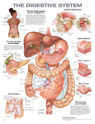
Original medical illustrations by Brian Evan in consultation with Mark Frasier, Professor of Anatomy, Colorado State University
Consultant: Nicole R. Herring, PhD