The
Eye Anatomical Chart, Second Edition features general anatomy of the eye with colorful detailed illustrations, all fully labeled.
- Outer eye with surface anatomy, anterior view, also showing the lacrimal gland and nasolacrimal duct
- Eyeball in skull, lateral and top views
- Visual field diagram
- Cross section of the eye, lateral view, as the large central illustration along with the anterior chamber angle
- Medial cross section of the eye
- The cornea, macula lutea, fundus, and retina in close-up views
20" x 26" heavy paper laminated
?Consultant (2025): Christopher J. Rapuano, MD, Wills Eye Hospital
Original medical illustrations by Keith Kasnot, CMI, in consultation with Randall Paul, OD, Phoenix, Arizona
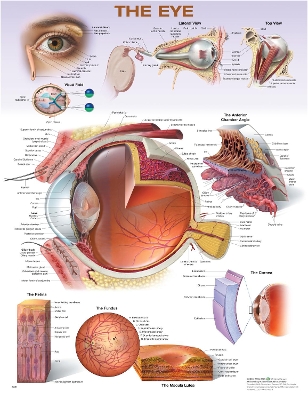
The
Eye Anatomical Chart, Second Edition features general anatomy of the eye with colorful detailed illustrations, all fully labeled.
- Outer eye with surface anatomy, anterior view, also showing the lacrimal gland and nasolacrimal duct
- Eyeball in skull, lateral and top views
- Visual field diagram
- Cross section of the eye, lateral view, as the large central illustration along with the anterior chamber angle
- Medial cross section of the eye
- The cornea, macula lutea, fundus, and retina in close-up views
20" x 26" heavy paper laminated
Consultant (2025): Christopher J. Rapuano, MD, Wills Eye Hospital
Original medical illustrations by Keith Kasnot, CMI, in consultation with Randall Paul, OD, Phoenix, Arizona