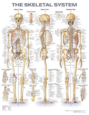
The Skeletal System, Second Edition is a modern update of the bestselling, classic The Skeletal System chart dating back to 1947. The illustrations are vividly colored and finely detailed, and structures are clearly labeled. The chart features three large illustrations showing the anterior, lateral, and posterior views of the skeletal system. Eight smaller illustrations show:
- Portion of Long Bone
- Auditory Ossicles (Left Medial View 3X)
- Ligaments of Right hand (Dorsal and Palmar Views)
- Ligaments of Right Foot (Dorsal and Plantar View)
- Right Knee Joint (Anterior and Posterior Views)
20" x 26" heavy paper laminated with grommets at top corners